CHAPTER 2
HUMAN REPRODUCTION
2.1 The Male Reproductive System
2.2 The Female Reproductive System
2.3 Gametogenesis
2.4 Menstrual Cycle
2.5 Fertilisation and Implantation
2.6 Pregnancy and Embryonic Development
2.7 Parturition and Lactation
It is commonly understood that humans engage in sexual reproduction and exhibit viviparity. The sequence of reproductive processes in humans encompasses the generation of gametes, specifically spermatozoa in males and ova in females (a process termed gametogenesis). Subsequent steps involve the introduction of spermatozoa into the female reproductive canal (insemination) and the subsequent union of male and female gametes (fertilisation), culminating in the formation of a zygote. Following this, a blastocyst forms and develops, attaching itself to the uterine lining (implantation). This leads to the progression of embryonic development, known as gestation, and ultimately, the birth of the infant (parturition). It has been established that these reproductive phenomena commence post-puberty. Notable distinctions exist in the reproductive timelines of males and females; for instance, spermatogenesis persists into advanced age in men, whereas oogenesis typically ceases in women around their fifth decade of life. We shall now proceed to investigate the human male and female reproductive systems.
2.1 : THE MALE REPRODUCTIVE SYSTEM
Situated within the pelvic area (Figure 2.1a), the male reproductive apparatus comprises a pair of testes, supplementary ducts, various glands, and the external genitalia.
The testes reside externally to the abdominal cavity, housed within a sac known as the scrotum. This scrotal positioning is crucial for thermoregulation, maintaining the testes at a temperature approximately 2–2.5°C below the typical internal body temperature, a condition vital for successful spermatogenesis. In mature individuals, each testis exhibits an oval morphology, typically measuring 4 to 5 cm in length and 2 to 3 cm in width. A robust outer layer encapsulates each testis, which is internally subdivided into approximately 250 distinct units termed testicular lobules (Figure 2.1b).
Within each lobule are found one to three extensively coiled seminiferous tubules, serving as the sites of sperm generation. The internal surface of each seminiferous tubule is populated by two distinct cell populations: male germ cells (spermatogonia) and Sertoli cells (Figure 2.2). The male germ cells undergo meiotic processes, culminating in the production of spermatozoa, whereas the Sertoli cells are responsible for providing nutritional support to these developing germ cells. The areas situated external to the seminiferous tubules, referred to as interstitial spaces, encompass fine blood vessels and specialized interstitial cells, also known as Leydig cells (Figure 2.2). These Leydig cells are instrumental in the biosynthesis and secretion of testicular hormones, specifically androgens. Additionally, other immune-responsive cells are present within these interstitial regions.
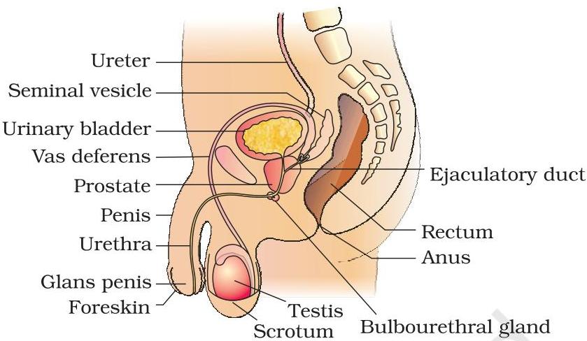 Figure 2.1(a) Diagrammatic sectional view of male pelvis showing reproductive system
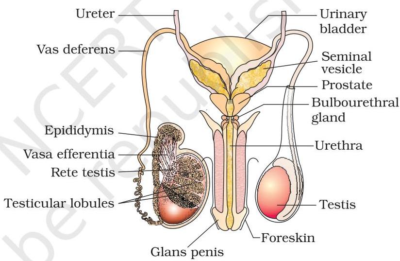 Figure 2.1(b) Diagrammatic view of male reproductive system (part of testis is open to show inner details)
The accessory ducts of the male reproductive system comprise the rete testis, vasa efferentia, epididymis, and vas deferens (Figure 2.1b). From the seminiferous tubules of the testis, spermatozoa pass into the vasa efferentia via the rete testis. The vasa efferentia then exit the testis and connect to the epididymis, which is positioned along the posterior aspect of each testis. The epididymis transitions into the vas deferens, a tube that ascends into the abdominal cavity and arches over the urinary bladder. This duct merges with a duct from the seminal vesicle, forming the ejaculatory duct, which subsequently empties into the urethra (Figure 2.1a). Collectively, these ducts serve to store and convey sperm from the testes for external release via the urethra. The urethra itself originates from the urinary bladder and traverses the length of the penis, terminating at its external orifice, the urethral meatus.
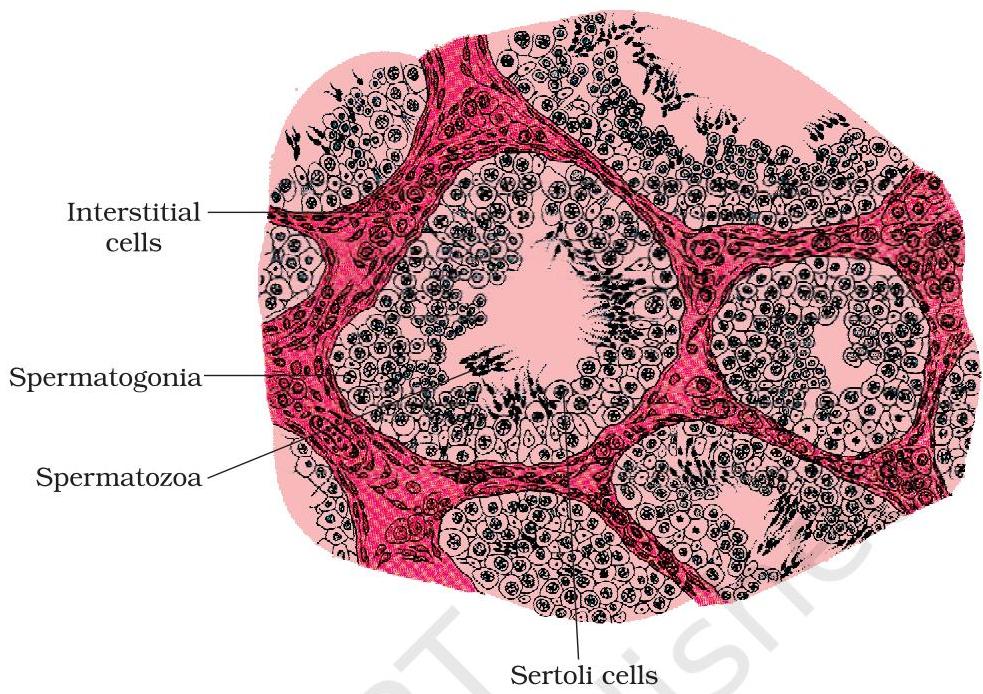 Figure 2.2 Diagrammatic sectional view of seminiferous tubule
The penis represents the external genitalia in males (Figure 2.1a, b). It consists of specialized erectile tissue that enables penile erection, a mechanism crucial for facilitating insemination. The distal, expanded portion of the penis is termed the glans penis, which is typically enveloped by a retractable fold of skin known as the foreskin.
The male accessory glands (Figure 2.1a, b) encompass paired seminal vesicles, a single prostate gland, and paired bulbourethral glands. The collective secretions from these glands form the seminal plasma, a fluid notable for its high concentrations of fructose, calcium ions, and various enzymes. Furthermore, the secretions originating from the bulbourethral glands contribute to the lubrication of the penis.
2.2 : THE FEMALE REPRODUCTIVE SYSTEM
Comprising a pair of ovaries, oviducts, the uterus, cervix, vagina, and external genitalia, the female reproductive system is situated within the pelvic region (Figure 2.3a). These components, in conjunction with the mammary glands, are structurally and functionally coordinated to facilitate ovulation, fertilisation, gestation, parturition, and postnatal care.
The ovaries serve as the principal female gonads, responsible for generating the female gamete (ovum) and synthesizing various steroid hormones, collectively known as ovarian hormones. Positioned bilaterally within the lower abdomen (Figure 2.3b), each ovary measures approximately 2 to 4 cm in length and is anchored to both the pelvic wall and the uterus via ligaments. A delicate epithelial layer encapsulates each ovary, encasing the ovarian stroma, which itself is compartmentalized into two distinct regions: an outer cortex and an internal medulla.
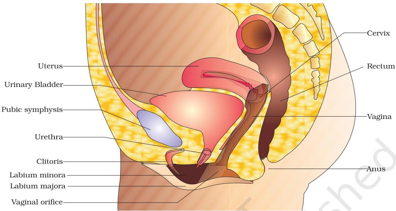 Figure 2.3 (a) Diagrammatic sectional view of female pelvis showing reproductive system
The oviducts, also known as fallopian tubes, along with the uterus and vagina, form the female accessory ducts. Each fallopian tube measures approximately $10 - 12\mathrm{cm}$ in length, originating near the ovarian periphery and extending towards the uterus (Figure 2.3b). The segment nearest the ovary is a funnel-shaped structure termed the infundibulum. Its margins are adorned with digit-like extensions called fimbriae, which are instrumental in capturing the ovum subsequent to ovulation. The infundibulum transitions into a broader
 Figure 2.3 (b) Diagrammatic sectional view of the female reproductive system
portion of the oviduct designated as the ampulla. The terminal segment of the oviduct, known as the isthmus, exhibits a constricted lumen and connects with the uterus.
The uterus, a singular organ often referred to as the womb, possesses a morphology resembling an inverted pear. It receives structural support from ligaments anchored to the pelvic wall. The uterus communicates with the vagina via a constricted region known as the cervix. The internal space of the cervix is termed the cervical canal (Figure 2.3b), which, together with the vagina, constitutes the birth canal. The uterine wall is composed of three distinct tissue layers: the outermost, delicate membranous perimetrium; the thick, intermediate layer of smooth muscle, the myometrium; and the innermost glandular layer, the endometrium, which lines the uterine cavity. The endometrium undergoes cyclic transformations throughout the menstrual cycle, whereas the myometrium generates powerful contractions during parturition.
The external female genitalia encompass the mons pubis, labia majora, labia minora, hymen, and clitoris (Figure 2.3a). The mons pubis is an adipose tissue cushion enveloped by skin and pubic hair. The labia majora consist of prominent fleshy tissue folds that descend from the mons pubis and enclose the vaginal orifice. Beneath the labia majora are the paired tissue folds known as the labia minora. The vaginal opening is frequently, though not always completely, occluded by a membranous structure termed the hymen. The clitoris is a diminutive, digitiform organ situated at the superior juncture of the two labia minora, positioned above the urethral meatus. While the hymen commonly ruptures during initial coitus (sexual intercourse), its integrity can also be compromised by sudden trauma such as a fall or jolt, the insertion of a vaginal tampon, or engagement in certain physical activities like horseback riding or cycling. Furthermore, in some individuals, the hymen may remain intact even after coitus. Consequently, the presence or absence of the hymen does not serve as a dependable indicator of virginity or prior sexual activity.
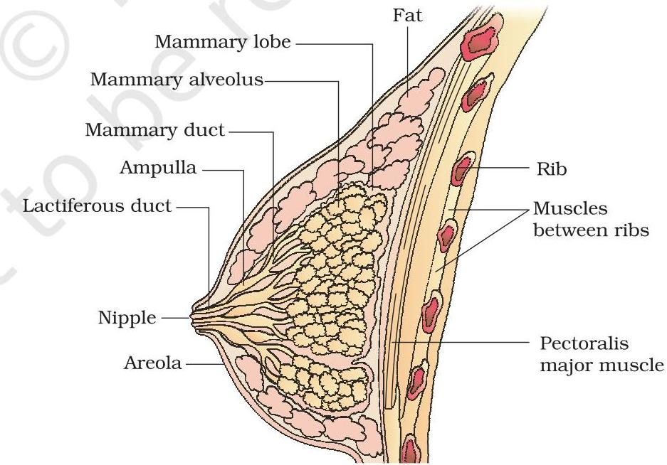 Figure 2.4 A diagrammatic sectional view of Mammary gland
HUMAN REPRODUCTION
A functional mammary gland is a characteristic feature of all female mammals. These paired structures, known as breasts, consist of glandular tissue along with varying amounts of adipose tissue. The glandular tissue within each breast is organized into 15-20 mammary lobes, which contain clusters of cells called alveoli (Figure 2.4). The cells lining the alveoli are responsible for secreting milk, which is then stored within the cavities (lumens) of these alveoli. The alveoli drain into mammary tubules. The tubules from each lobe converge to form a mammary duct. Several mammary ducts then coalesce to create a broader mammary ampulla, which is connected to the lactiferous duct and opens at the nipple, facilitating the expression of milk during lactation.
2.3 : GAMETOGENESIS
The primary reproductive organs—the testes in males and the ovaries in females—generate gametes (sperms and ova, respectively) through a process termed gametogenesis. In the testes, immature male germ cells (spermatogonia) produce sperms via spermatogenesis, a process that commences at puberty. The spermatogonia (singular: spermatogonium), located on the inner wall of the seminiferous tubules, proliferate by mitotic division, thereby increasing their numbers. Each spermatogonium is diploid, containing 46 chromosomes. A subset of these spermatogonia, designated as primary spermatocytes, periodically undergoes meiosis. A primary spermatocyte completes the first meiotic division (reduction division), resulting in the formation of two equal, haploid cells known as secondary spermatocytes, each possessing only 23 chromosomes. These secondary spermatocytes then proceed through the second meiotic division to generate four equal, haploid spermatids (Figure 2.5). What would be the number of chromosomes in the spermatids? Subsequently, spermatids transform into spermatozoa (sperms) through a process called spermiogenesis. Following spermiogenesis, the heads of the spermatozoa become embedded within Sertoli cells, and are ultimately released from the seminiferous tubules by a process termed spermiation.
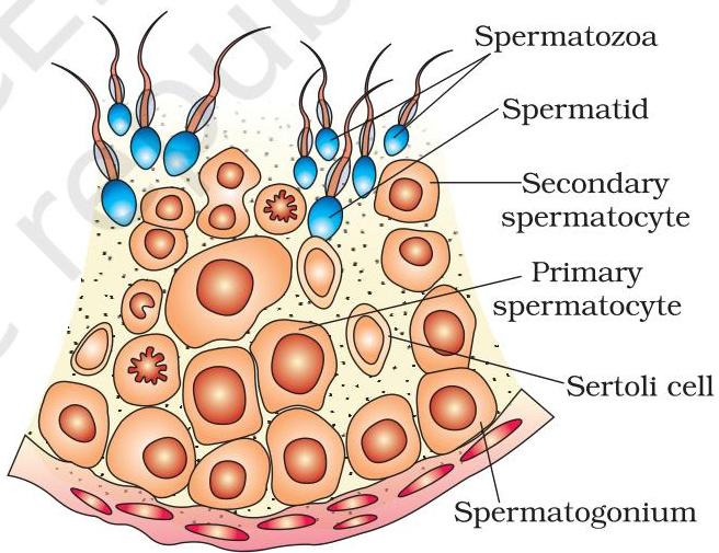 Figure 2.5 Diagrammatic sectional view of a seminiferous tubule (enlarged)
Spermatogenesis is initiated at puberty, driven by a significant increase in the secretion of gonadotropin-releasing hormone (GnRH). This hormone, as you may recall, originates from the hypothalamus. Elevated levels of GnRH subsequently act upon the anterior pituitary gland, stimulating the release of two gonadotropins: luteinizing hormone (LH) and follicle-stimulating hormone (FSH). LH primarily targets Leydig cells, promoting the synthesis and secretion of androgens. These androgens, in turn, stimulate the process of spermatogenesis. Concurrently, FSH acts on Sertoli cells and stimulates
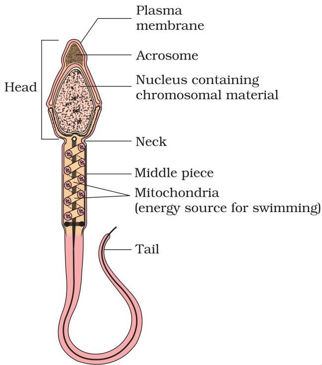 Figure 2.6 Structure of a sperm
Certain secreted factors contribute to the process of spermiogenesis.
A microscopic examination of a sperm reveals its distinct structural components: a head, a neck, a middle piece, and a tail (Figure 2.6). The entirety of the sperm's anatomy is encased by a plasma membrane. Within the sperm head lies an elongated, haploid nucleus, whose anterior region is surmounted by a cap-like formation known as the acrosome. The acrosome is replete with enzymes instrumental in facilitating the fertilisation of the ovum. The middle piece is characterized by the presence of numerous mitochondria, which are responsible for generating the energy required for tail movement, thereby enabling sperm motility—a critical prerequisite for successful fertilisation. During a single coitus, a human male typically ejaculates approximately 200 to 300 million spermatozoa. For optimal fertility, it is stipulated that at least 60 percent of these spermatozoa must exhibit normal morphology and size, and at least 40 percent must demonstrate vigorous motility.
Following their release from the seminiferous tubules, spermatozoa undergo transport via the male accessory ducts. The secretions emanating from the epididymis, vas deferens, seminal vesicle, and prostate gland are indispensable for the maturation and motility of the spermatozoa. The collective mixture of seminal plasma and spermatozoa constitutes the semen. The functional integrity of the male sex accessory ducts and glands is sustained by testicular hormones, specifically androgens.
The developmental pathway leading to the formation of a mature female gamete is termed oogenesis, a process notably distinct from spermatogenesis. Oogenesis commences during the embryonic developmental phase when several million gamete mother cells, or oogonia, are generated within each fetal ovary; crucially, no additional oogonia are formed or added subsequent to birth. These progenitor cells initiate division and enter prophase-I of meiosis, where they become temporarily arrested, at which point they are designated as primary oocytes. Subsequently, each primary oocyte becomes enveloped by a layer of granulosa cells, forming what is known as a primary follicle (Figure 2.7). A substantial proportion of these follicles undergo degeneration between birth and puberty. Consequently, by the time puberty is reached, only 60,000 to 80,000 primary follicles remain in each ovary. As development progresses, primary follicles acquire additional layers of granulosa cells and a newly formed theca, thereby transforming into secondary follicles.
The secondary follicle soon undergoes a transformation into a tertiary follicle, which is distinctly characterized by the presence of a fluid-filled cavity known as the antrum. The theca layer differentiates into an internal stratum, the theca interna, and an external stratum, the theca externa. It is noteworthy that at this particular stage, the primary oocyte residing within the tertiary follicle experiences an increase in size and completes its first meiotic division. This division is unequal, yielding a large, haploid secondary oocyte and a diminutive first polar body (Figure 2.8b). The
secondary oocyte retains the majority of the nutrient-rich cytoplasm from the primary oocyte. Can you identify any adaptive advantage for this unequal distribution? Does the first polar body, generated from the first meiotic division, undergo further division or does it degenerate? At present, a definitive answer regarding its fate remains uncertain. The tertiary follicle further matures into the Graafian follicle, also recognized as the mature follicle (Figure 2.7). Concurrently, the secondary oocyte develops a new surrounding membrane called the zona pellucida. The Graafian follicle then ruptures, culminating in the release of the secondary oocyte (ovum) from the ovary, a process termed ovulation. Can you identify major differences between
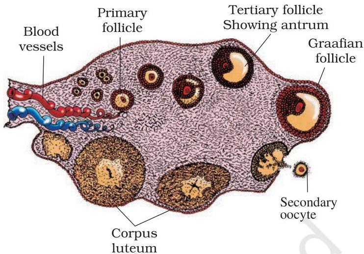 Figure 2.7 Diagrammatic Section view of ovary
Regarding spermatogenesis and oogenesis, a diagrammatic depiction of these processes is provided below (Figure 2.8).
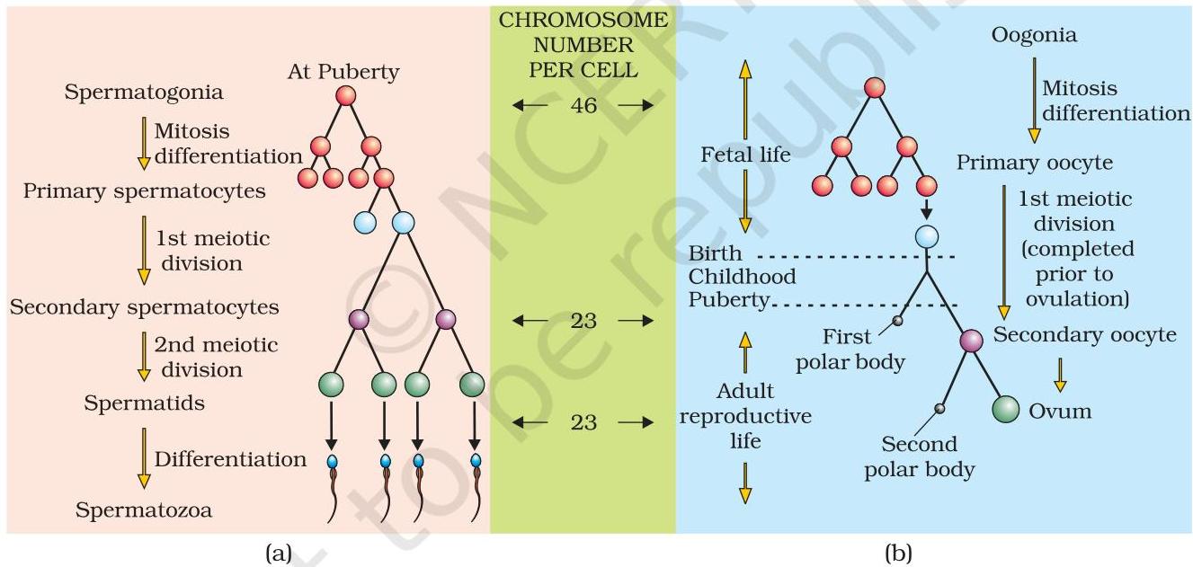 Figure 2.8 Schematic representation of (a) Spermatogenesis; (b) Oogenesis
2.4 : MENSTRUAL CYCLE
The reproductive cycle observed in female primates (e.g., monkeys, apes, and human beings) is designated as the menstrual cycle. The initial onset of menstruation, occurring at puberty, is referred to as menarche. In human females, menstruation recurs at an approximate interval of 28 to 29 days, and the sequence of physiological events spanning from one menstruation to the subsequent one constitutes the menstrual cycle. Typically, a single ovum is discharged (ovulation) around the midpoint
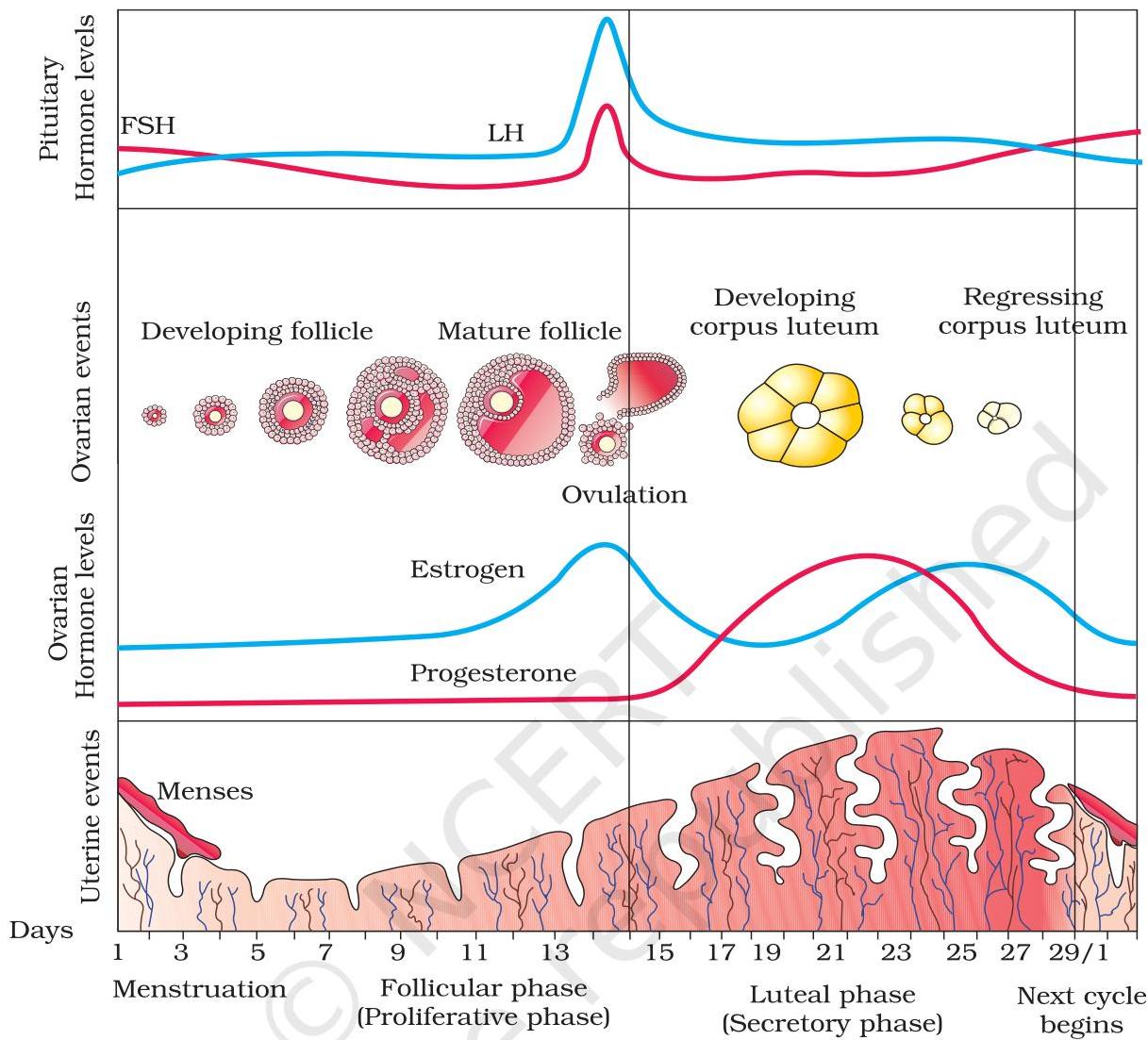 Figure 2.9 Diagrammatic presentation of various events during a menstrual cycle
of each menstrual cycle. The primary events of the menstrual cycle are delineated in Figure 2.9. The cycle initiates with the menstrual phase, during which menstrual flow occurs, lasting for 3 to 5 days. This menstrual discharge results from the breakdown of the uterine endometrial lining and its associated blood vessels, forming a fluid that exits through the vagina. Menstruation exclusively takes place if the released ovum has not been fertilised. An absence of menstruation can signify pregnancy; however, it may also be attributed to other underlying factors such as stress or compromised health. The menstrual phase is succeeded by the follicular phase. During this period, the primary follicles within the ovary develop into a fully mature Graafian follicle, while concurrently, the uterine endometrium regenerates through proliferative processes. These transformations in the ovary and uterus are regulated by fluctuations in the concentrations of pituitary and ovarian hormones (Figure 2.9). The secretion of
gonadotropins (LH and FSH) gradually increases throughout the follicular phase, stimulating both follicular development and the production of estrogens by the maturing follicles. Both LH and FSH attain their peak levels in the middle of the cycle (around the $14^{\text{th}}$ day). A rapid release of LH, reaching its maximum concentration during the mid-cycle, known as the LH surge, precipitates the rupture of the Graafian follicle and, consequently, the expulsion of the ovum (ovulation). Ovulation (the ovulatory phase) is succeeded by the luteal phase, during which the residual components of the Graafian follicle differentiate into the corpus luteum (Figure 2.9). The corpus luteum secretes substantial quantities of progesterone, a hormone crucial for the maintenance of the endometrium. Such a prepared endometrium is indispensable for the implantation of a fertilised ovum and for other processes associated with pregnancy. Throughout gestation, all events of the menstrual cycle cease, and menstruation does not occur. In the absence of fertilisation, the corpus luteum degenerates. This degeneration leads to the disintegration of the endometrium, resulting in menstruation and thereby marking the commencement of a new cycle. In humans, menstrual cycles typically conclude around 50 years of age, a phenomenon termed menopause. Cyclic menstruation serves as an indicator of a normal reproductive phase, extending from menarche to menopause.
2.5 : FERTILISATION AND IMPLANTATION
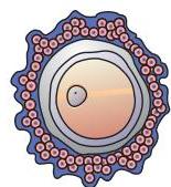
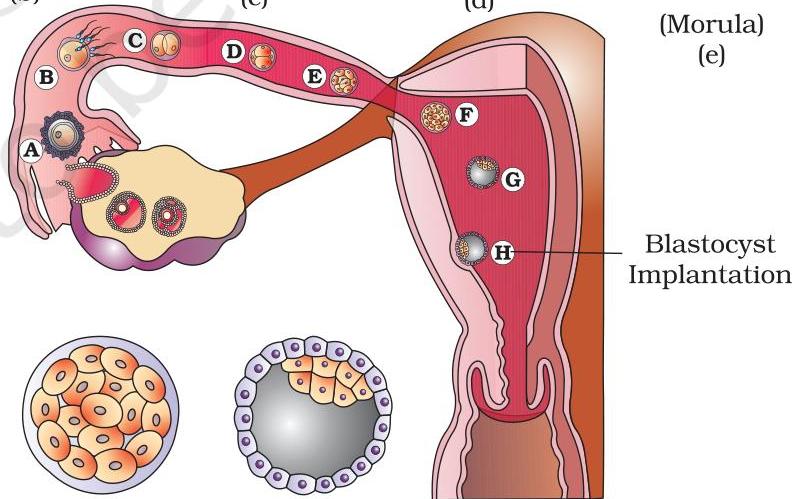
Sexual intercourse (coitus) involves the release of seminal fluid from the penis into the vaginal canal, a process termed insemination. Subsequently, the highly motile spermatozoa navigate rapidly through the cervix, traverse the uterine cavity, and ultimately reach the ampullary segment of the fallopian tube (Figure 2.11b). Concurrently, the oocyte released from the ovary is also transported to this same ampullary region, establishing the location for fertilisation. For fertilisation to transpire, the synchronous arrival of both the ovum and spermatozoa in the ampullary region is imperative. This critical requirement explains why not every instance of copulation culminates in fertilisation and subsequent pregnancy.
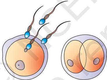
Fertilisation is defined as the process entailing the fusion of a spermatozoon with an oocyte. During this event, a sperm initially makes contact with the zona pellucida, an outer layer surrounding the ovum (Figure 2.10). This interaction triggers modifications within the oocyte's membrane, which serve to prevent the entry of additional spermatozoa. Consequently, this mechanism ensures that only a single sperm fertilises an ovum. The enzymatic secretions housed within the acrosome of the sperm facilitate its penetration through the zona pellucida and the plasma membrane, allowing it to enter the cytoplasm of the ovum.
Menstrual Hygiene
The maintenance of proper hygiene and sanitation practices during menstruation is of paramount importance. Individuals should bathe and cleanse themselves regularly. It is advised to use either sanitary napkins or meticulously cleaned homemade pads. These sanitary products, whether commercial or homemade, must be changed approximately every 4-5 hours, or as required by individual needs. Used sanitary napkins should be disposed of responsibly by wrapping them securely in a piece of used paper. Under no circumstances should used napkins be discarded into toilet drainpipes or left exposed in open areas. Following the handling of any used sanitary material, hands must be thoroughly washed with soap.
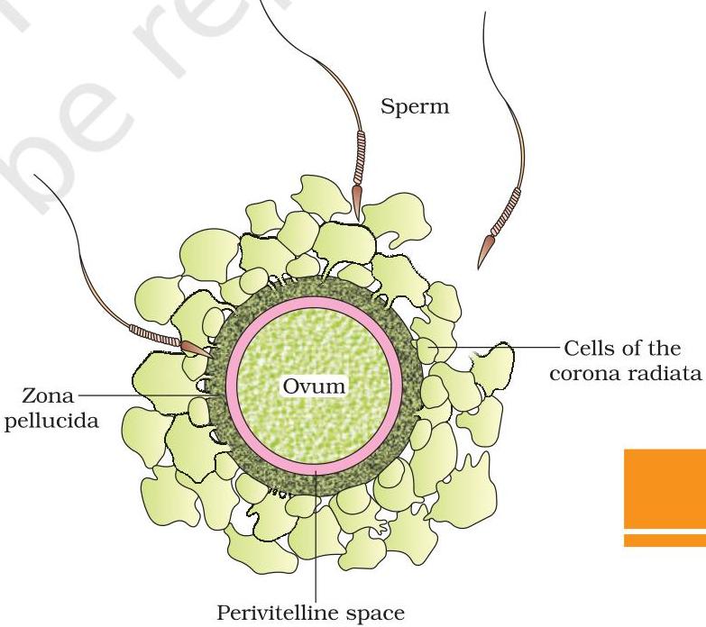 Figure 2.10 Ovum surrounded by few sperms
Interaction with the ovum's membrane triggers the completion of the secondary oocyte's meiotic division. This second meiotic division is also asymmetrical, resulting in the formation of a second polar body and a haploid ovum (ootid). Subsequently, the haploid nuclei of the sperm and the ovum coalesce to form a diploid zygote. What would be the chromosomal count within this zygote?
It is crucial to recognize that the biological sex of the offspring is determined at this very early stage. To understand this mechanism, consider the human chromosomal patterns: females possess an XX configuration, while males exhibit an XY pattern. Consequently, all haploid gametes (ova) produced by a female invariably carry an X sex chromosome. In contrast, male gametes (sperms) can carry either an X or a Y sex chromosome, with approximately 50 percent of sperms containing an X chromosome and the remaining 50 percent carrying a Y chromosome. Upon the fusion of male and female gametes, the resulting zygote will acquire either an XX or an XY chromosomal complement, depending on whether an X-carrying or a Y-carrying sperm fertilised the ovum. A zygote with an XX constitution will develop into a female infant, whereas an XY zygote will form a male (further details on chromosomal patterns are provided in Chapter 5). Hence, it is scientifically accurate to state that the sex of the baby is determined by the father, not by the mother.
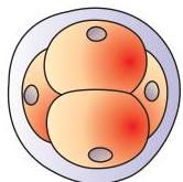
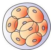
As the zygote transits through the isthmus region of the oviduct towards the uterus, it commences a series of mitotic divisions, a process known as cleavage (Figure 2.11). This process generates 2, 4, 8, and ultimately 16 daughter cells, which are termed blastomeres. An embryonic structure composed of 8 to 16
 (a)
(a)
 (b)
(b)
 (d)
(d)
 (Morula) (e)
 (f)
(Blastocyst) (g)
Blastocyst Implantation
Figure 2.11 Transport of ovum, fertilisation and passage of growing embryo through fallopian tube
(f)
(Blastocyst) (g)
Blastocyst Implantation
Figure 2.11 Transport of ovum, fertilisation and passage of growing embryo through fallopian tube
blastomeres is referred to as a morula (Figure 2.11e). The morula continues its mitotic divisions and undergoes transformation into a blastocyst (Figure 2.11g) as it progresses further into the uterine cavity. Within the blastocyst, the blastomeres organize into an external layer known as the trophoblast and an internal cluster of cells, termed the inner cell mass, which is connected to the trophoblast. Subsequently, the trophoblast layer establishes attachment to the endometrium, while the inner cell mass differentiates to form the embryo proper. Following this attachment, the uterine cells proliferate rapidly, enveloping the blastocyst. Consequently, the blastocyst becomes embedded within the uterine endometrium (Figure 2.11, step H). This process is termed implantation, and it marks the initiation of pregnancy.
2.6 : PREGNANCY AND EMBRYONIC DEVELOPMENT
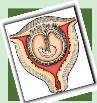
Subsequent to implantation, the trophoblast develops digit-like extensions known as chorionic villi. These villi are encompassed by the maternal blood and uterine tissue. A reciprocal interlocking occurs between the chorionic villi and the uterine tissue, culminating in the formation of the placenta (Figure 2.12), which serves as the crucial structural and functional interface connecting the developing embryo (fetus) and the maternal organism.
The placenta performs vital functions by facilitating the transfer of oxygen and essential nutrients to the embryo, while simultaneously eliminating carbon dioxide and metabolic waste products generated by the embryo. This connection between the placenta and the embryo is established via the umbilical cord, which acts as a conduit for the bidirectional transport of these substances. Beyond its transport capabilities, the placenta functions as an endocrine organ, synthesizing various hormones such as human chorionic gonadotropin (hCG), human placental lactogen (hPL), estrogens, and progestogens. Furthermore, during the later stages of gestation, the ovaries secrete relaxin. It is noteworthy that hCG, hPL, and relaxin are exclusively produced in females during pregnancy. Concurrently, maternal blood levels of other hormones, including estrogens, progestogens, cortisol, prolactin, and thyroxine, experience a multifold elevation throughout pregnancy. This surge in hormone production is crucial for sustaining fetal development, inducing necessary metabolic adaptations in the mother, and ensuring the successful maintenance of the pregnancy.
Immediately after implantation, the inner cell mass (embryo) differentiates
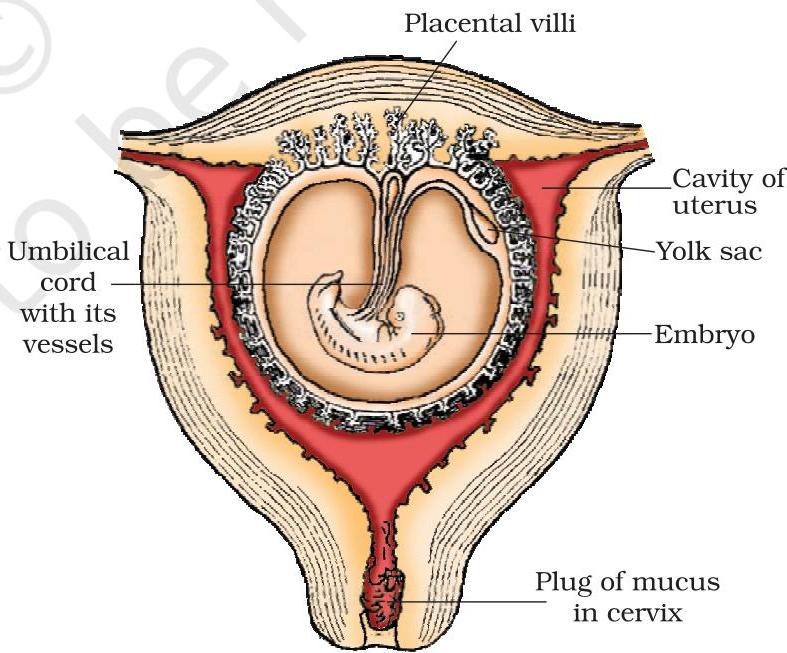 Figure 2.12 The human foetus within the uterus
Subsequent to implantation, the inner cell mass, which constitutes the embryo, undergoes differentiation to form two primary germ layers: an external layer termed the ectoderm and an internal layer termed the endoderm. Shortly thereafter, a third layer, the mesoderm, emerges positioned between the ectoderm and endoderm. These three embryonic germ layers are the progenitors of all tissues and organs found in the adult organism. It is pertinent to note that the inner cell mass comprises specialized cells known as stem cells, possessing the remarkable capacity to differentiate into all types of tissues and organs.
What are the significant developmental characteristics observed during different stages of pregnancy? Human gestation typically extends for a period of nine months. (Consider researching the gestational periods of other mammals such as dogs, elephants, and cats.) In human development, the embryonic heart begins to form after one month of gestation. A primary indication of fetal growth can be detected by carefully auscultating the heart sounds using a stethoscope. By the conclusion of the second month of pregnancy, the fetus exhibits the development of limbs and digits. The end of 12 weeks, marking the close of the first trimester, sees the formation of most major organ systems, with structures like the limbs and external genital organs becoming well-defined. Fetal movements and the initial appearance of hair on the head are commonly observed around the fifth month. Approaching the end of approximately 24 weeks (the conclusion of the second trimester), the fetal body is covered with fine hair, the eyelids separate, and eyelashes develop. Finally, by the end of the nine-month gestational period, the fetus achieves full maturation, preparing for parturition.
2.7 : PARTURITION AND LACTATION
Human gestation typically spans approximately nine months, a duration referred to as the gestation period. The expulsion or delivery of the fetus, driven by powerful uterine contractions as pregnancy concludes, constitutes a process known as parturition (childbirth). This intricate event is orchestrated by a sophisticated neuroendocrine mechanism. The initial stimuli for parturition arise from the fully matured fetus and the placenta, prompting gentle uterine contractions termed the fetal ejection reflex. This reflex, in turn, stimulates the maternal pituitary gland to release oxytocin. Oxytocin then acts upon the uterine musculature, intensifying contractions and thereby promoting additional oxytocin secretion. This positive feedback loop, where uterine contractions stimulate further oxytocin release, progressively escalates the strength of these contractions. Ultimately, this leads to the infant's expulsion from the uterus via the birth canal, completing parturition. Subsequently, the placenta is also expelled from the uterus shortly after the baby's delivery. What substance do physicians commonly administer to initiate labor?
During pregnancy, the female mammary glands differentiate, commencing milk production nearing the end of gestation through a process termed lactation. This physiological function enables the mother to nourish the newborn infant. The milk secreted in the initial days of lactation is known as colostrum, characterized by its rich content of antibodies that are critically important for establishing immunity in neonates. Medical professionals advocate for breastfeeding during the early stages of infant development to foster optimal health.
SUMMARY
Human beings engage in sexual reproduction and are viviparous. The male reproductive apparatus comprises a pair of testes, along with the male sex accessory ducts, accessory glands, and external genitalia. Within each testis, approximately 250 distinct compartments, known as testicular lobules, are present. Each of these lobules typically houses one to three extensively coiled seminiferous tubules. The internal lining of each seminiferous tubule consists of spermatogonia and Sertoli cells. Spermatogonia undergo meiotic processes to generate spermatozoa, whereas Sertoli cells furnish essential nourishment to these developing germ cells. Positioned outside the seminiferous tubules, Leydig cells are responsible for synthesizing and secreting testicular hormones, specifically androgens. The external genital structure in males is termed the penis.
The female reproductive apparatus encompasses a pair of ovaries, a pair of oviducts, the uterus, the vagina, external genitalia, and a pair of mammary glands. The ovaries serve to generate the female gamete, the ovum, alongside various steroid hormones, collectively known as ovarian hormones. Within the ovarian stroma, follicles at diverse developmental stages are embedded. The oviducts, uterus, and vagina collectively constitute the female accessory ducts. The uterine wall is composed of three distinct layers: the perimetrium, myometrium, and endometrium. The female external genitalia comprises the mons pubis, labia majora, labia minora, hymen, and clitoris. Mammary glands are recognized as a key female secondary sexual characteristic.
Spermatogenesis culminates in the production of spermatozoa, which are subsequently conveyed through the male sex accessory ducts. A typical human spermatozoon is structurally delineated into a head, a neck, a middle piece, and a tail. The process responsible for the genesis of mature female gametes is termed oogenesis. In female primates, the reproductive cycle is known as the menstrual cycle, which commences upon the attainment of sexual maturity, or puberty. During each menstrual cycle, typically only a single ovum is released during ovulation. The cyclic transformations observed in the ovary and uterus throughout the menstrual cycle are orchestrated by fluctuating levels of pituitary and ovarian hormones. Following coitus, spermatozoa are conveyed to the ampulla, where a sperm fertilises the ovum, resulting in the formation of a diploid zygote. The chromosomal contribution from the sperm, specifically the presence of an X or Y chromosome, dictates the sex of the developing embryo. The zygote then undergoes successive mitotic divisions to form a blastocyst, which subsequently implants into the uterine wall, initiating pregnancy. After a gestational period of approximately nine months, the fully developed fetus is prepared for birth. The process of childbirth, termed parturition, is orchestrated by an intricate neuroendocrine mechanism involving cortisol, estrogens, and oxytocin. Mammary glands undergo differentiation during pregnancy and commence milk secretion post-childbirth. The newborn infant is nourished with maternal milk (lactation) during its initial months of development.
EXERCISES
- Fill in the blanks: (a) Human beings exhibit reproduction through a __________ (asexual/sexual) mechanism. (b) Humans are classified as __________ (egg-laying, live-bearing, egg-retaining-then-live-bearing) organisms. (c) In Homo sapiens, the process of fertilisation occurs __________ (externally/internally). (d) The reproductive cells (gametes) from males and females are __________ (diploid/haploid). (e) The resultant zygote is __________ (diploid/haploid).
(f) The phenomenon involving the expulsion of an ovum from a fully developed ovarian follicle is termed __________ . (g) The hormonal agent responsible for triggering ovulation is designated as __________ . (h) The amalgamation of male and female reproductive cells is referred to as __________ . (i) The site of fertilisation is __________ . (j) The zygote undergoes cellular division to generate ___________, which subsequently implants within the uterine wall. (k) The anatomical entity establishing a vascular conduit between the developing fetus and the maternal uterus is known as __________ .
Illustrate, with appropriate labels, the male reproductive system.
Provide a labelled illustration of the female reproductive system.
Enumerate two primary functions for both the testis and the ovary.
Detail the structural composition of a seminiferous tubule.
Define spermatogenesis. Provide a concise description of the spermatogenic process.
Identify the hormones implicated in the regulatory control of spermatogenesis.
Provide definitions for spermiogenesis and spermiation.
Present a labelled diagram depicting a spermatozoon.
Specify the principal constituents of seminal plasma.
Outline the primary physiological roles of the male accessory ducts and glands.
What constitutes oogenesis? Furnish a brief explanation of the oogenetic process.
Produce a labelled illustration representing a cross-section of an ovary.
Illustrate, with labels, a Graafian follicle.
State the functions associated with each of the subsequent entities: (a) Corpus luteum
(b) Endometrium
(c) Acrosome
(d) Sperm tail
(e) FimbriaeDetermine the veracity (True/False) of the following statements. For each false assertion, revise it to render it accurate. (a) Androgens are synthesized by Sertoli cells. (True/False)
(b) Spermatozoa derive nourishment from Sertoli cells. (True/False)
(c) Leydig cells are located within the ovary. (True/False)
(d) Leydig cells are responsible for androgen synthesis. (True/False)
(e) Oogenesis transpires within the corpus luteum. (True/False)
(f) The menstrual cycle terminates during gestation. (True/False)
(g) The existence or non-existence of the hymen does not serve as a dependable gauge of virginity or prior sexual activity. (True/False)Define the menstrual cycle. Which hormonal agents govern its regulation?
Explain parturition. Which hormones participate in the initiation of parturition?
Within many societal contexts, females are frequently held accountable for the birth of female offspring. Elucidate why this attribution is inaccurate.
What is the typical number of oocytes released by a human ovary over a one-month period? If a mother delivered monozygotic (identical) twins, how many oocytes would you infer were released? Would your response differ if the twins born were dizygotic (fraternal)?
Estimate the number of oocytes released by the ovary of a female canine that produced a litter of six puppies.