UNIT 5
HUMAN PHYSIOLOGY
Chapter 14: Breathing and Exchange of Gases
Chapter 15: Body Fluids and Circulation
Chapter 16: Excretory Products and their Elimination
Chapter 17: Locomotion and Movement
Chapter 18: Neural Control and Coordination
Chapter 19: Chemical Coordination and Integration
The reductionist approach to study of life forms resulted in increasing use of physico-chemical concepts and techniques. Majority of these studies employed either surviving tissue model or straightaway cellfree systems. An explosion of knowledge resulted in molecular biology. Molecular physiology became almost synonymous with biochemistry and biophysics. However, it is now being increasingly realised that neither a purely organismic approach nor a purely reductionistic molecular approach would reveal the truth about biological processes or living phenomena. Systems biology makes us believe that all living phenomena are emergent properties due to interaction among components of the system under study. Regulatory network of molecules, supramolecular assemblies, cells, tissues, organisms and indeed, populations and communities, each create emergent properties. In the chapters under this unit, major human physiological processes like, exchange of gases, blood circulation, locomotion and movement are described in cellular and molecular terms. The last two chapters point to the coordination and regulation of body events at the organismic level.
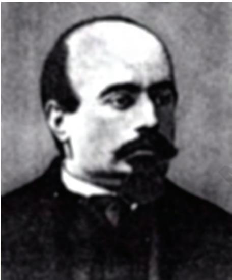
Alfonso Corti (1822-1888)
Alfonso Corti, Italian anatomist, was born in 1822. Corti began his scientific career studying the cardiovascular systems of reptiles. Later, he turned his attention to the mammalian auditory system. In 1851, he published a paper describing a structure located on the basilar membrane of the cochlea containing hair cells that convert sound vibrations into nerve impulses, the organ of Corti. He died in the year 1888.
CHAPTER 14
BREATHING AND EXCHANGE OF GASES
14.1 Respiratory Organs
14.2 Mechanism of Breathing
14.3 Exchange of Gases
14.4 Transport of Gases
14.5 Regulation of Respiration
14.6 Disorders of Respiratory System
As you have read earlier, oxygen $\mathrm{O_2}$ is utilised by the organisms to indirectly break down simple molecules like glucose, amino acids, fatty acids, etc., to derive energy to perform various activities. Carbon dioxide ($\mathrm{CO_2}$) which is harmful is also released during the above catabolic reactions. It is, therefore, evident that $\mathrm{O_2}$ has to be continuously provided to the cells and $\mathrm{CO_2}$ produced by the cells have to be released out. This process of exchange of $\mathrm{O_2}$ from the atmosphere with $\mathrm{CO_2}$ produced by the cells is called breathing, commonly known as respiration. Place your hands on your chest; you can feel the chest moving up and down. You know that it is due to breathing. How do we breathe? The respiratory organs and the mechanism of breathing are described in the following sections of this chapter.
14.1 RESPIRATORY ORGANS
Mechanisms of breathing vary among different groups of animals depending mainly on their habitats and levels of organisation. Lower invertebrates like sponges, coelenterates, flatworms, etc., exchange $\mathrm{O_2}$ with $\mathrm{CO_2}$ by simple diffusion over their entire body surface. Earthworms use their moist cuticle and insects have a network of tubes (tracheal tubes) to transport atmospheric air within the body. Special vascularised structures called gills (branchial respiration) are used by most of the aquatic arthropods and molluscs whereas vascularised bags called lungs (pulmonary respiration) are used by the terrestrial forms for the exchange of gases. Among vertebrates, fishes use gills whereas amphibians, reptiles, birds and mammals respire through lungs. Amphibians like frogs can respire through their moist skin (cutaneous respiration) also.
14.1.1 Human Respiratory System
We have a pair of external nostrils opening out above the upper lips. It leads to a nasal chamber through the nasal passage. The nasal chamber opens into the pharynx, a portion of which is the common passage for food and air. The pharynx opens through the larynx region into the trachea. Larynx is a cartilaginous box which helps in sound production and hence called the sound box. During swallowing glottis can be covered by a thin elastic cartilaginous flap called epiglottis to prevent the entry of food into the larynx. Trachea is a straight tube extending up to the mid-thoracic cavity, which divides at the level of 5th thoracic vertebra into a right and left primary bronchi. The trachea, primary, secondary and tertiary bronchi, and initial bronchioles are supported by incomplete cartilaginous rings. Each terminal bronchiole gives rise to a number of very thin, irregular-walled and vascularised bag-like structures called alveoli. The branching network of bronchi, bronchioles and alveoli comprise the lungs (Figure 14.1). We have two lungs which are covered by a double layered pleura, with pleural fluid between them. It reduces friction on the lung-surface. The outer pleural membrane is in close contact with the thoracic
lining whereas the inner pleural membrane is in contact with the lung surface. The part starting with the external nostrils up to the terminal bronchioles constitute the conducting part whereas the alveoli and their ducts form the respiratory or exchange part of the respiratory system. The conducting part transports the atmospheric air to the alveoli, clears it from foreign particles, humidifies and also brings the air to body temperature. Exchange part is the site of actual diffusion of $\mathrm{O_2}$ and $\mathrm{CO_2}$ between blood and atmospheric air.
 Figure 14.1 Diagrammatic view of human respiratory system (sectional view of the left lung is also shown)
The lungs are situated in the thoracic chamber which is anatomically an air-tight chamber. The thoracic chamber is formed dorsally by the vertebral column, ventrally by the sternum, laterally by the ribs and on the lower side by the dome-shaped diaphragm. The anatomical setup of lungs in thorax is such that any change in the volume of the thoracic cavity will be reflected in the lung (pulmonary) cavity. Such an arrangement is essential for breathing, as we cannot directly alter the pulmonary volume.
Respiration involves the following steps:
(i) Breathing or pulmonary ventilation by which atmospheric air is drawn in and $\mathrm{CO_2}$ rich alveolar air is released out. (ii) Diffusion of gases ($\mathrm{O_2}$ and $\mathrm{CO_2}$) across alveolar membrane. (iii) Transport of gases by the blood. (iv) Diffusion of $\mathrm{O_2}$ and $\mathrm{CO_2}$ between blood and tissues. (v) Utilisation of $\mathrm{O_2}$ by the cells for catabolic reactions and resultant release of $\mathrm{CO_2}$ (cellular respiration as dealt in the Chapter 12).
14.2 MECHANISM OF BREATHING
Breathing involves two stages: inspiration during which atmospheric air is drawn in and expiration by which the alveolar air is released out. The movement of air into and out of the lungs is carried out by creating a pressure gradient between the lungs and the atmosphere. Inspiration can occur if the pressure within the lungs (intra-pulmonary pressure) is less than the atmospheric pressure, i.e., there is a negative pressure in the lungs with respect to atmospheric pressure. Similarly, expiration takes place when the intra-pulmonary pressure is higher than the atmospheric pressure. The diaphragm and a specialised set of muscles – external and internal intercostals between the ribs, help in generation of such gradients. Inspiration is initiated by the contraction of diaphragm which increases the volume of thoracic chamber in the antero-posterior axis. The contraction of external inter-costal muscles lifts up the ribs and the
sternum causing an increase in the volume of the thoracic chamber in the dorso-ventral axis. The overall increase in the thoracic volume causes a similar increase in pulmonary volume. An increase in pulmonary volume decreases the intrapulmonary pressure to less than the atmospheric pressure which forces the air from outside to move into the lungs, i.e., inspiration (Figure 14.2a). Relaxation of the diaphragm and the inter-costal muscles returns the diaphragm and sternum to their normal positions and reduce the thoracic volume and thereby the pulmonary volume. This leads to an increase in intra-pulmonary pressure to slightly above the atmospheric pressure causing the expulsion of air from the lungs, i.e., expiration (Figure 14.2b). We have the ability to increase the strength of inspiration and expiration with the help of additional muscles in the abdomen. On an average, a healthy human breathes 12-16 times/minute. The volume of air involved in breathing movements can be estimated by using a spirometer which helps in clinical assessment of pulmonary functions.
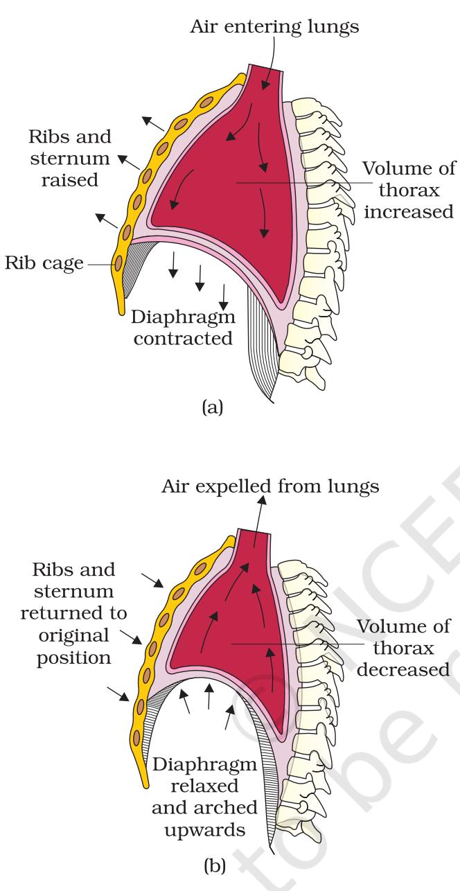 Figure 14.2 Mechanism of breathing showing : (a) inspiration (b) expiration
14.2.1 Respiratory Volumes and Capacities
Tidal Volume (TV): Volume of air inspired or expired during a normal respiration. It is approx. $500~\mathrm{mL}$, i.e., a healthy man can inspire or expire approximately 6000 to 8000 mL of air per minute.
Inspiratory Reserve Volume (IRV): A person can inspire by a forcible inspiration. This averages $2500\mathrm{mL}$ to $3000\mathrm{mL}$.
Expiratory Reserve Volume (ERV): A person can expire by a forcible expiration. This averages $1000\mathrm{mL}$ to $1100\mathrm{mL}$.
Residual Volume (RV): Volume of air remaining in the lungs even after a forcible expiration. This averages $1100\mathrm{mL}$ to $1200\mathrm{mL}$.
By adding up a few respiratory volumes described above, one can derive various pulmonary capacities, which can be used in clinical diagnosis.
Inspiratory Capacity (IC): Total volume of air a person can inspire after a normal expiration. This includes tidal volume and inspiratory reserve volume ($\mathrm{TV+IRV}$).
Expiratory Capacity (EC): Total volume of air a person can expire after a normal inspiration. This includes tidal volume and expiratory reserve volume ($\mathrm{TV+ERV}$).
Functional Residual Capacity (FRC): Volume of air that will remain in the lungs after a normal expiration. This includes ERV+RV.
Vital Capacity (VC): The maximum volume of air a person can breathe in after a forced expiration. This includes ERV, TV and IRV or the maximum volume of air a person can breathe out after a forced inspiration.
Total Lung Capacity (TLC): Total volume of air accommodated in the lungs at the end of a forced inspiration.